68 Male patient k/c/o of HTN with slurring of speech
This is an online E log book to discuss our patient's de-identified health data shared after taking his/her/guardian's signed informed consent.
Here we discuss our individual patient's problems through series of inputs from available global online community of experts with an aim to solve those patient's clinical problems with collective current best evidence based inputs.
This E log book also reflects my patient-Centered online learning portfolio and your valuable inputs on the comment box is welcome."
CHEIF COMPLAINTS
68 year old male patient resident of deverakonda Telangana farmer by occupation , uneducated .
Right handed individual
Informant - son
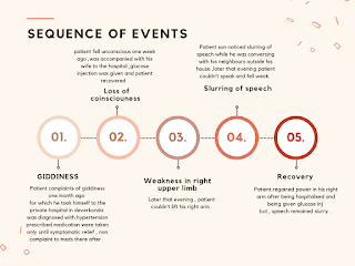
- Giddiness since one month
- Weakness of right upper limb 3 days ago
- Slurring of speech since 3 days ago
sequence of events
- No history of trauma, fever
- No history of headache, vomiting
- No history of loss of consciousness or seizures
- No history of decreased smell, blurring of vision, double vision, decreased sensation over face, decreased hearing or vertigo, difficulty in swallowing or nasal regurgitation on swallowing and no hoarseness of voice
- No history of tremors
- No history of chest pain or palpitation
- hypertensive elderly man with sudden onset, progressive Paralysis of right sided upper limb ,unable to comment on sensation over face due to associated aphasia.
- History is suggestive of of acute neurological deficit probably due to ischemic stroke involving left MCA territory
General Physical Examination
- A elderly man who is moderately build and nourished is conscious and cooperative and cannot comment orientation due to aphasia
- Patient is right handed
- Pallor: Not seen
- Icterus: Not seen
- Cyanosis: Not seen
- Clubbing: Not seen
- Lymphadenopathy: Not seen
- Pedal Edema: Not seen
Vitals
- Pulse: 82 beats per min, regular, normal in volume and character
- Respiratory rate: 18 cycles/ min, abdominothoracic type
- BP: 160/94 mm of Hg in left brachial artery on day of admission
- On day of examination-130/70mm of Hg
- Temperature: 98.6 degree Farenheit
Higher Mental Functions
- Right handed individual
- Conscious and cooperative, unable to comment on orientation to time place and person due to aphasia
- Appearance and Behavior: appropriate.
- Patient is emotionally stable
- Calculation: unable to comment
- Speech: Speech fluency markedly reduced. Incoherent speech, Agrammatic speech ,Repetition is absent
- Comprehension present but delayed .
- Patient is uneducated hence cannot comment on reading and follow
flexion : 5/5 5/5 https://youtube.com/shorts/W1WHEit_N4k?feature=share
Extension 5/5. 5/5
Abduction 5/5. 5/5
Adduction 5/5. 5/5
Internal rotation 5/5. 5/5
External rotation 5/5. 5/5
Elbow:
Flexion. 5/5. 5/5
Extension:5/5. 5/5
Wrist:
Flexion:5/5. 5/5
Extension:5/5. 5/5
Abduction : 5/5. 5/5
adduction:5/5. 5/5
HIP
Posterior column: Fine touch, Vibration, Position sense present on all limbs
1. Spontaneous speech
2. Comprehension- Intact able to point out four objects
Yes or no response - is this a hospital ? He said yes
Is this ur wife ? Yes
has ur speech impaired -yes
Do u know any songs ? -yes
Can u sing ? -yes
u named someone just a while ago whose name was that ? Was it ur wife ?? -yes ( tired to say bharya)
Complex command - patient cannot articulate a proper sentence
3. Repetition -absent
4. Naming and Word finding-
Due to aphasia cannot be assessed
5.Reading - patient had never received education
6.Writing –
Cerebellar Functions
- Nystagmus: Not seen
- Dysmetria/past pointing: Not seen
- Intentional tremors: not seen
- Dysdiadokinesia: not seen
- Gait: normal
- No signs of meningeal irritation
- Examination of skull and spine is normal
- Ausculation of neck: no carotid bruit heard
PER ABDOMEN EXAMNATION
-Scaphoid
-No visible pulsations/engorged veins/sinuses
-Soft,non tender, no guarding and rigidity, no organomegaly
-Bowel sounds heard
SYSTEMIC EXAMINATION
CVS
Elliptical & bilaterally symmetrical chest
-No visible pulsations/engorged veins on the chest
-Apex beat seen in 5th intercostal space medial to mid clavicular line
-S1 S2 heard
-No murmurs
RESPIRATORY SYSTEM
Upper respiratory tract normal
Lower respiratory tract :
-Trachea is central
-Movements are equal on both sides
-On percussion resonant on all areas
-Bilateral air entry equal
-Normal vesicular breath sounds heard
-No added sounds
-Vocal resonance equal on both sides in all areas
INVESTIGATIONS
CT SCAN OF BRAIN PLAIN
FINDINGS:
Ischaemic changes noted in bilateral gangliocapsular region.
Small focal chronic lacunar infarcts noted in right side thalamus and left side basal ganglia.
Age related atrophy.
Rest of cerebral parenchyma shows normal gray / white matter differentiation.
Posterior fossa structures including fourth ventricle are normal.
Supratentorial ventricular system is normal.
Cortical sulci, sylvian fissures and basal cisterns are normal.
No midline shift.
No extra axial collection.
IMPRESSION:
• MILD CHRONIC CEREBRAL SMALL VESSELS ISCHAEMIC DISEASE CHANGES WITH SMALL FOCAL CHRONIC LACUNAR INFARCTS AS DESCRIBED ABOVE.
Bilirubin 0.81
D. Bilirubin 0 . 20
A/G Ratio 1.36
PROVISIONAL DIAGNOSIS
Acute onset neurological deficit in the form of right upper limb paresis with involvement of left hemisphere , probably involving MCA territory , with involvement of speech in the form of broca’s aphasia ,along with infarct in B/L gangliocapsular area with chronic infraction in Rt thalamus and left basal ganglia probable etiology of HTN and DM
https://www.ncbi.nlm.nih.gov/books/NBK563216/
CT scan seldom identifies lacunar ischemic insult within the first 24 hours due to its small size. If seen, lacunar strokes are ill-defined hypodensities on CT scans unless there is a hemorrhagic component to the acute stroke. A hyperdensity of a large artery on non-contrast head CT indicates the presence of a thrombus inside the arterial lumen or vessel calcification. Early infarct signs on non-contrast CT include loss of gray-white differentiation and focal hypoattenuation of brain parenchyma. These details are difficult to read in small subcortical strokes. Chronic lesions may appear as hypodense foci.


Comments
Post a Comment